In the realm of tiny details, where the minutiae of the natural world come into clear view, understanding how to effectively use precision optical instruments becomes essential. This guide aims to provide a comprehensive overview of operating these sophisticated devices, ensuring users can fully appreciate the intricate wonders that lie hidden from the naked eye.
From setting up the equipment to mastering its features, this document will walk you through every necessary step. Whether you’re a beginner or looking to refine your skills, knowing the correct procedures will enhance your experience and improve your observations.
By delving into the functionality and maintenance of these devices, you’ll gain valuable insights and practical knowledge. This will not only aid in accurate and effective usage but also foster a deeper appreciation for the complexities of microscopic exploration.
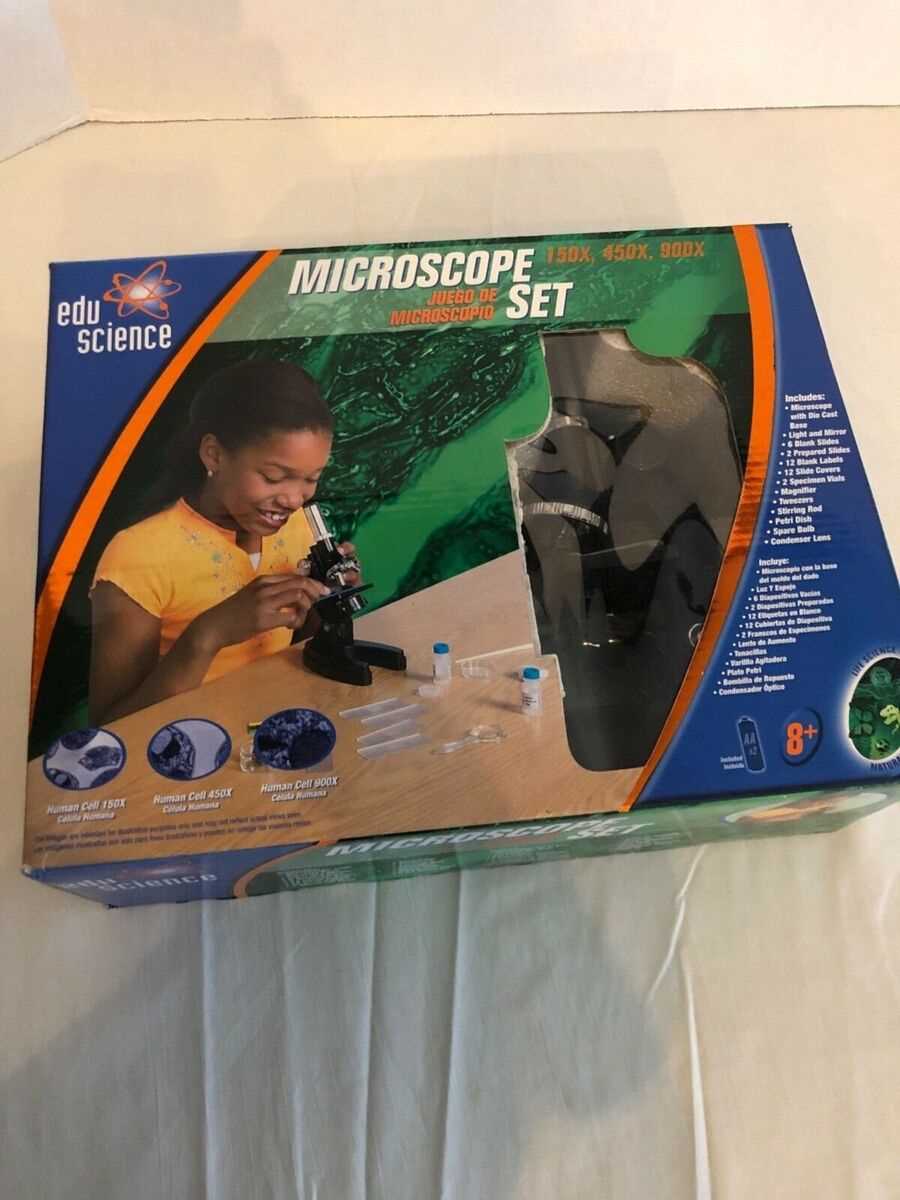
Understanding Your Edu Science Microscope
To get the most out of your optical instrument, it’s essential to grasp its fundamental components and their functions. This section provides a comprehensive overview of the key elements and features that contribute to the effective use of your device. Familiarity with these parts will enhance your ability to explore and analyze samples with precision.
Firstly, it is important to recognize the primary sections of your tool, including the base, arm, and stage. Each part plays a crucial role in the stability and adjustment of your observations. The base provides support and stability, while the arm connects the base to the optical system. The stage holds the specimen in place and allows for precise movement and positioning.
Additionally, the optical elements such as the objective lenses and eyepiece are vital for magnification and clarity. Understanding how to adjust these components ensures that you can achieve the desired level of detail and resolution. Proper alignment and focus adjustments are key to obtaining clear and accurate images.
Finally, becoming acquainted with the adjustment knobs and their functions will facilitate smooth operation. These knobs allow you to control the focus and illumination of your observations, ensuring that you can adapt to various sample types and lighting conditions.
By familiarizing yourself with these aspects, you will be well-equipped to utilize your optical instrument effectively and make the most of its capabilities.
Essential Components of Your Microscope
Understanding the core elements of your optical device is crucial for effective use and maintenance. Each part plays a significant role in magnifying and analyzing specimens, ensuring you achieve accurate and clear observations. Familiarizing yourself with these components will enhance your overall experience and proficiency.
Key Elements

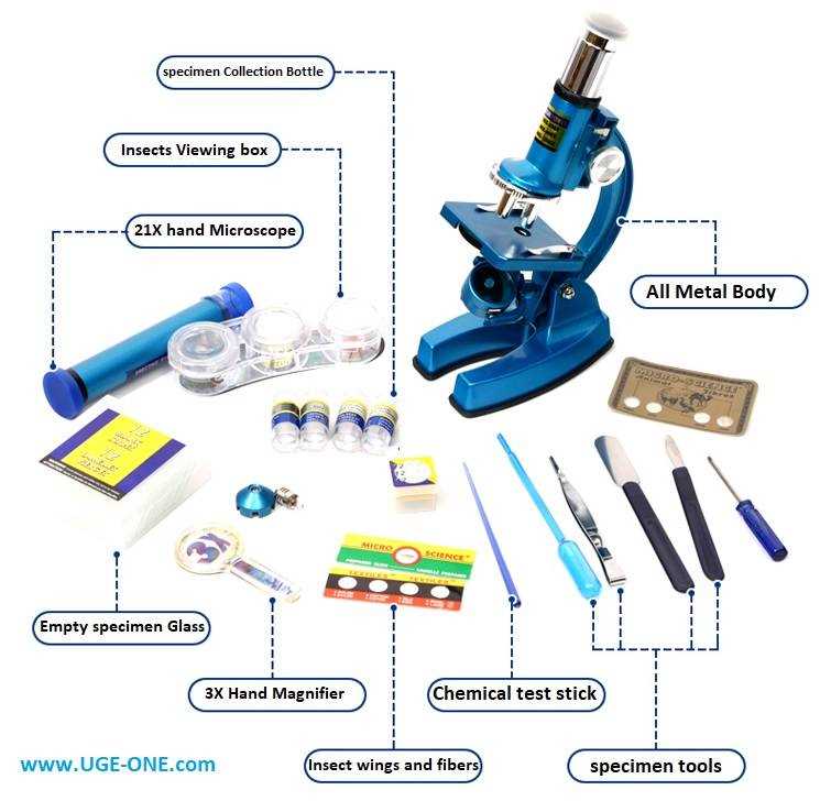
- Optical System: This includes the lenses that magnify the image, such as the ocular lens and objective lenses. They are essential for viewing and focusing on the sample.
- Illumination: The light source, usually positioned at the base, provides the necessary brightness for clear visibility of the specimen.
- Stage: The platform where the sample is placed. It often includes mechanisms for adjusting the position of the slide.
- Focus Mechanism: Controls that allow you to bring the specimen into sharp view. This typically includes coarse and fine adjustment knobs.
- Arm and Base: The structure that supports and stabilizes the device, providing a sturdy base and a handle for easy manipulation.
Additional Features
- Condenser: Positioned beneath the stage, it focuses light onto the specimen to improve image clarity.
- Diaphragm: Controls the amount of light reaching the specimen, aiding in contrast and resolution.
- Stage Clips: Hold the slide in place, preventing movement during observation.
Setting Up the Microscope Correctly
To ensure optimal performance and accurate observations, it is essential to prepare your optical instrument properly before use. This preparation involves several key steps that guarantee the equipment is ready for detailed examination and analysis. By following these guidelines, you can maximize the effectiveness of your observations and maintain the longevity of the device.
First, place the instrument on a stable, flat surface to prevent any movement or vibrations that could affect the clarity of your observations. Ensure that the workspace is clean and free from any debris that might obstruct the view or damage the delicate components.
Next, adjust the focus mechanism to the lowest magnification setting. This allows for easier alignment and prevents accidental damage to the sample or the lens system. Carefully check the alignment of the lenses and make sure that each optical component is securely in place.
Additionally, ensure that the light source is properly positioned and adjusted. The illumination should be even and bright enough to provide clear visibility of the sample. If your device includes a condenser, adjust it according to the sample’s requirements to enhance the lighting quality and contrast.
Finally, verify that all moving parts, such as the stage and focus knobs, are functioning smoothly. Any resistance or irregularity might indicate the need for maintenance or adjustment. By ensuring everything is operating correctly, you set the stage for accurate and effective examination.
By carefully following these steps, you set up your optical instrument for success, ensuring precise and reliable results during your observations.
Proper Use of Optical Settings
Achieving clear and precise observations through your optical instrument relies significantly on the correct adjustment of its visual parameters. Proper management of these settings ensures that the images you see are sharp, detailed, and accurately represent the subject under examination. Understanding how to fine-tune these elements is crucial for obtaining high-quality results and making the most of your observational tasks.
To begin, familiarize yourself with the various adjustments available, such as focus, magnification, and illumination. Each component plays a vital role in the overall clarity and quality of the image. Start by setting up the initial focus to bring the specimen into view, then gradually enhance the magnification for finer details. Adjust the light source to achieve optimal brightness and contrast, which will further refine the visibility of intricate features.
Regular calibration and careful handling of these settings not only improve accuracy but also extend the longevity of your equipment. Always ensure that adjustments are made gently and in small increments to avoid any potential damage or misalignment. By mastering these adjustments, you will enhance your observational capabilities and achieve the best possible results from your optical instrument.
Cleaning and Maintenance Tips
Proper upkeep of your optical equipment ensures its longevity and optimal performance. Regular cleaning and maintenance are essential to prevent dirt, debris, and other contaminants from affecting the quality of your observations. Here, you will find key practices to keep your device in excellent condition.
Routine Cleaning Procedures
To maintain clarity and precision, clean the lenses and optical surfaces with care. Use a soft brush or air blower to remove loose dust particles. For more thorough cleaning, apply a few drops of lens cleaning solution to a lint-free cloth and gently wipe the surfaces in a circular motion. Avoid using paper products or rough materials that could scratch the optics.
Maintenance Practices
Regularly check all mechanical components for smooth operation. Ensure that the moving parts are free from obstructions and that any lubrication is applied according to the manufacturer’s recommendations. Store your equipment in a dry, dust-free environment to prevent internal moisture and dirt buildup. Additionally, periodically inspect and calibrate your instrument to ensure its accuracy and functionality.
Common Troubleshooting Solutions
In any technical setup, encountering issues is a common occurrence. Understanding how to address and resolve these problems efficiently is crucial for maintaining optimal performance. This section provides practical steps to diagnose and fix typical issues that may arise.
- Issue: Poor Image Quality
- Check the alignment of the optical components to ensure they are properly positioned.
- Verify that the lens surfaces are clean and free from dust or smudges.
- Adjust the focus and illumination settings to enhance clarity.
- Issue: Difficulty in Viewing Samples
- Ensure that the sample is properly mounted and secured.
- Confirm that the appropriate objective lens is in use for the sample type.
- Adjust the stage and focus knobs to achieve a clear view.
- Issue: Mechanical Movement Problems
- Inspect moving parts for any signs of obstruction or damage.
- Lubricate moving components if necessary, using the recommended products.
- Check for any loose screws or parts and tighten them as needed.
- Issue: Lighting Issues
- Ensure that the light source is functioning and properly aligned.
- Check the intensity settings and adjust them to the desired level.
- Inspect the light path for any blockages or irregularities.
By following these steps, many common issues can be resolved efficiently. Regular maintenance and careful handling can further help in preventing these problems from recurring.
Advanced Features and Techniques

Exploring the sophisticated functionalities of optical devices can significantly enhance your observational capabilities. This segment delves into the intricate aspects that elevate your ability to analyze and interpret minute details with greater precision. Understanding and utilizing these advanced attributes will allow you to push the boundaries of your research and observation.
Digital Imaging Integration provides a modern approach to capturing and analyzing detailed visuals. Incorporating high-resolution cameras and software tools can facilitate the documentation and manipulation of images, allowing for enhanced clarity and detailed examination.
Phase Contrast is a technique used to observe transparent specimens without staining. By exploiting differences in the refractive index of the sample and the surrounding medium, you can achieve high-contrast images of live cells and other delicate structures.
Fluorescence Microscopy enables the visualization of specific components within a sample through the use of fluorescent dyes or tags. This method highlights particular structures or processes, providing valuable insights into cellular functions and molecular interactions.
High-Resolution Objectives offer improved magnification and clarity, allowing for the observation of finer details that might be missed with standard lenses. Employing these high-power objectives can significantly improve the resolution and quality of your images.
Mastering these advanced features requires practice and an understanding of their underlying principles. By integrating these techniques into your observational practices, you can achieve more detailed and accurate analyses of your samples.